September 19, 2024
Histologic Impacts Of A New Gadget For High-intensity Focused Ultrasound Cyclocoagulation Arvo Journals
Extracorporeal High-intensity Concentrated Ultrasound Therapy For Bust Cancer Cells Clinical Oncology And Cancer Research Study Secondly, in the HSI technique, we just utilized the noticeable variety for measurement. A more comprehensive wavelength array, such as 400-- 1,000 nm, may supply even more spooky information for improved assessment of healing result. The current wavelength array is limited by the hyperspectral imager used, which has a discovery array from 400 to 700 nm. Considering that the classification result of the HSI technique in the visible wavelength variety is not ideal, we will perform HSI information purchase in a wider wavelength range to supply a fair contrast in between the HSI and ADT methods in the future Cryotherapy study.Histological Evaluation Of Neuroinflammatory Feedbacks After Hifu Pulse-train Exposure
The datasets produced for this study are available on demand to the equivalent author. Where Iv( x, y, λ) is the reflection intensity of the skin in band λ at factor (x, y), and Iboard( x, y, λ) is the representation intensity of a PTFE board covering the skin in band λ at point (x, y). There are 2 means of getting the dark frames for reflectance adjustment. One is to maintain the lens covered during direct exposure [54] and the other is to switch off the source of light [48]High-intensity focused ultrasound for endometrial ablation in adenomyosis: a clinical study - Frontiers
High-intensity focused ultrasound for endometrial ablation in adenomyosis: a clinical study.
Posted: Tue, 25 Jun 2024 00:07:27 GMT [source]
Techniques
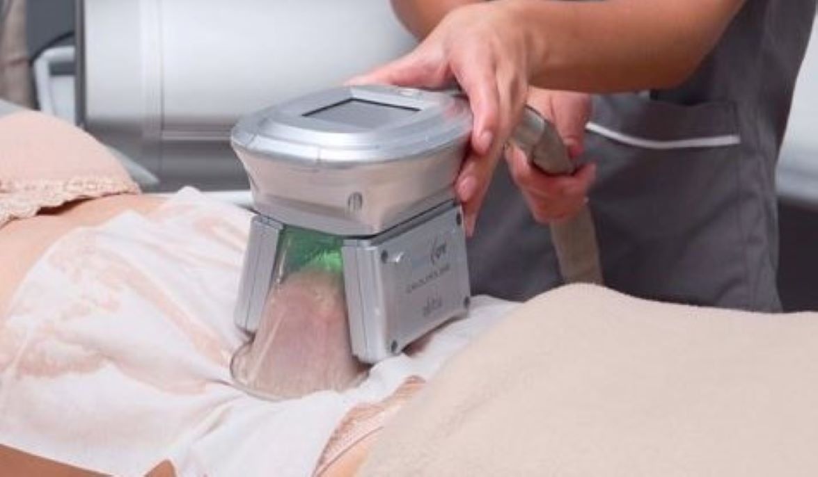
All rights are scheduled, consisting of those for message and data mining, AI training, and similar modern technologies. For all open access web content, the Creative Commons licensing terms use. This work was moneyed by Fda (FDA) Medical Countermeasures Initiative (MCMi) and laboratory fund of Department of Biomedical Physics at the Office of Science and Design Laboratories of the FDA. We would like to say thanks to Shaheda Mehtus for the assistance in histological staining, Lacey Walker for behavioral screening, and Dr. Bipasa Biswas for input on statistics.Early And Lasting Results Of Stomach Fat Reduction Using Ultrasound And Radiofrequency Therapies
There were no records of skin burns during subsequent medical researches, and no postmarketing records of skin burns have taken place in the European Union or Canada. To define patient outcomes following visually guided high-intensity focused ultrasound (HIFU) for focal treatment of local prostate cancer. To assess the clinical safety and security and efficiency of using highintensity concentrated ultrasound (HIFU) therapy, for bust cancer cells, and to choose the ideal methods in reviewing the restorative effects. Information of 55 clients with uterine fibroids (typical age 43.4 years, Table 1) treated with US-guided HIFU were analyzed. 3 reported major AE events (anemia, appendicitis, and pulmonary thromboembolism) were identified by the investigator to be unassociated to treatment with the HIFU device. The seriousness of the most common AE is summarized in Table 4 and defined in greater detail listed below. Treated and without treatment adipose tissue is shown in a low-magnification histology slide, with skin noticeable at the top of the photo. The well-demarcated thermal sore can be clearly recognized in the middle of the slide.- Complying with treatment, there was gross proof of mild cells ecchymosis from capillary damages.
- Every patient's problems, consisting of itching and skin lesion look, were reported, and RGB pictures of the vulva were caught by colposcopy before and after HIFU therapy as clinical regimens.
- No significant modifications in lab worths were observed in any type of studies, consisting of scientific chemistry and hematology parameters, serum lipids (Number 3), and liver function examinations.
- According to the particular optimals of the spectra of the treated locations before and after HIFU therapy, oxy-hemoglobin( HbO2), deoxy-hemoglobin( Hb), and methemoglobin (mHb) were utilized as the three major absorption components for further analysis.
- Temperature level data from the thermocouples were tape-recorded every 500 nanoseconds throughout of the treatment and for numerous seconds afterward.
- The authors recognize Irene Zender for her constant assistance in scientific management of HIFU-treated clients as well as Guido Luechters who supplied a useful assistance and advice in all statistical questions any time.
What is the success rate of HIFU?
In 2013 and 2014, 3 scientific studies (1;2;3) from French and German facilities published lasting results (one decade) of overall HIFU treatment and confirmed its effectiveness: according to cancer danger, certain survival rate at 10 years is in between 92% and 99%; survival price without transition at 10 years is in between 86% and ...
Social Links